Quantitative phase imaging of biological specimens using DHM

Digital Holographic Microscopy
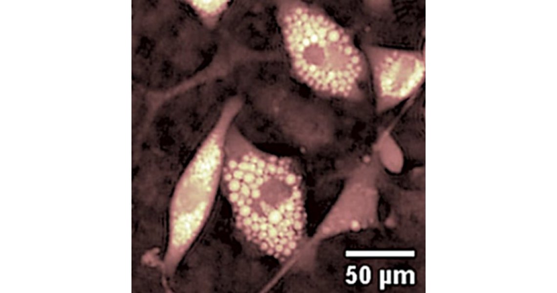
(DHM) uses one or more lasers to deliver quantitative phase imaging (QPI) of
biological specimens. By measuring optical path length changes referenced to
the background, it is possible to observe proteins and cells at high-resolution
without affecting them with bright light, dyes, fluorophores or other
preparations that could cause changes to physiological processes or motility.
A key feature of DHM is its ability to image the specimen at different depths without the need to refocus. This is also advantageous for continuous observations of live cells in 3D environments with various stages of confluence.
The phase contrast imaging
afforded by a DHM system can be used to determine cellular volume and water
content, intracellular solute concentrations, tissue density, cell thickness
and dry mass, making DHM an excellent label-free cytometry tool. Identification
and characterisation of cancer, blood and stem cells is thus made quicker and
easier, and time-lapse imaging of in vitro toxicity and drug testing
experiments simpler.
Lyncée Tec offer a number of different DHM systems for a range of biomedical applications. These include cell biology, cancer research, nanotoxicology and histopathology where the measurement of cellular end points or continuous label-free monitoring of growth and morphology changes are required.
For more information please contact us, visit the Lyncée Tec product pages, or read through the application notes listed below.


