ISS PL1 Microscopes


Overview
Benefits include :
-
FLIM of Large Areas
Upright Microscope: 100 mm x 100 mm
Inverted Microscope: 120 mm x 75 mm -
Lifetime Measurements
From 100 ps to 100 ms
-
Modularity
A selection of laser wavelengths, detectors, number of detection channels, & microscopes.
A time-resolved confocal microscope, the PL1 is designed primarily for material sciences research requiring the ultimate sensitivity in FLIM acquisition of large area samples. The sample, with dimensions up to 100 x 100 mm for the upright microscope and up to 120 x 75 mm for the inverted type, is placed on the high-precision, computer-controlled, XY stage that travels from pixel to pixel featuring a 22 nm resolution.
The excitation source is a laser diode, a pulsed laser, or a multiphoton laser. The fluorescence is collected by one detector covering the range 350 – 1050 nm; additional detectors can be added including a spectrograph for pixel spectral acquisition.
 PL1.png)
PL1Material Sciences Confocal Workstation
The Data is Clear!
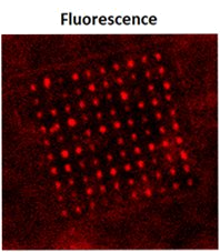
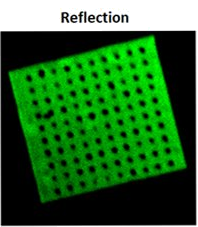
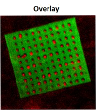
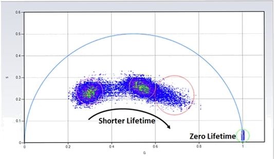
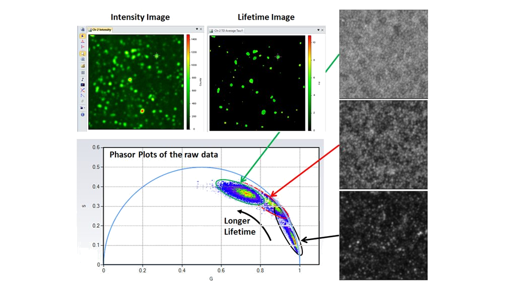
Phasor plots separate different lifetime species of quantum dots coated on the substrate directly from the raw data. Excitation was 470 nm, emission: 499 - 632 nm (fluorescence), 475/35 nm (reflection), scanning area 17.5 µm x 17.5 µm.
(Courtesy of Wenjie Liu and Dr. Yaowau Hu; Purdue University; West Lafayette, IN; USA)
Perovskite confocal lifetime imaging for both photoluminescence intensity and lifetime at the high spatial resolution. ISS VistaVision provides both the minimization fitting algorithm and the phasor plots for lifetime analysis; for example, using the multi-image phasor analysis routine, the lifetime changes in different Perovskite samples can be immediately identified by the phasor plots of their raw data sets (no fitting required), making the lifetime analysis much easier and robust. Excitation was 488 nm; emission through a 635 nm long pass filter; scanning area 20 µm x 20 µm.
(Courtesy of Dr. Xiaodan Zhang; Nan Kai University, Tianjin, P.R. China)
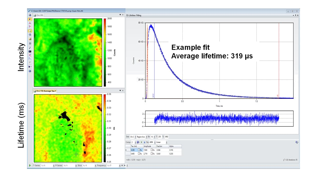
Upconversion nanoparticle confocal lifetime imaging for both photoluminescence intensity and lifetime at the high spatial resolution. Excitation: 980 nm, Emission: 509 - 552 nm, scanning area 20 µm x 20 µm
(Courtesy of Dr. Yan Guan, Peking University, Beijing; P.R. China)
| File Name | Size | Link |
|---|---|---|
|
|
207.42KiB | Download |


