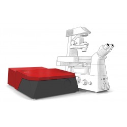
Aquila
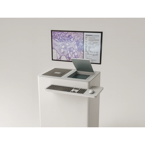

Please note: the Aquila is currently a research instrument, and should not be used as a medical device, but as a supplementary imaging technique for clinical research.
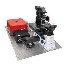
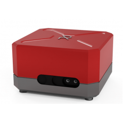
Aquila- Histological imaging at the point of care: taking SRS and chemical imaging out of the laser lab and moving into the Clinical environment- Fast and accurate cancer diagnostics.
Aqila combines proprietary laser imaging with workflow-ready design for next-generation tissue insights.
- Large field of view up to several centimeters
- Submicron spatial resolution
- 3D optical sectioning through several tens of micrometers
- Real time preview and rapid image acquisition
- Non-destructive imaging for intact downstream analysis
- Minimal tissue preparation — no stains or sectioning required
- Digital output for remote review and AI integration
Features
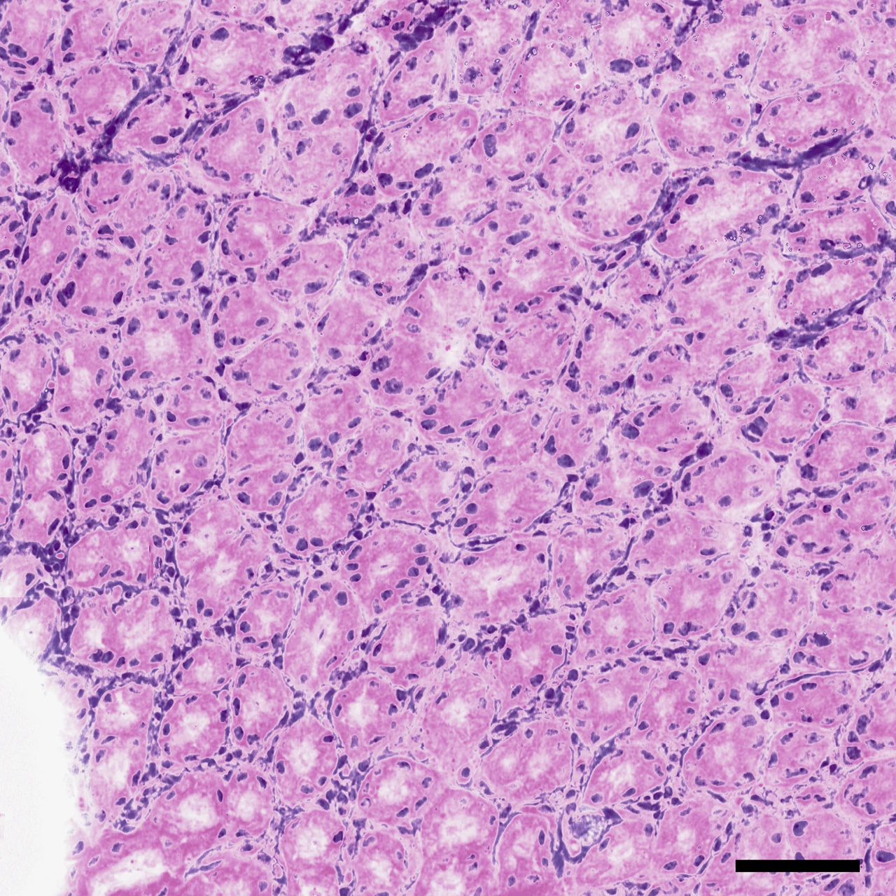
From overview to subcellular detail: Provides macro-to-micro visualization and multimodal optical signals in a single dataset.
 |
 |
 |
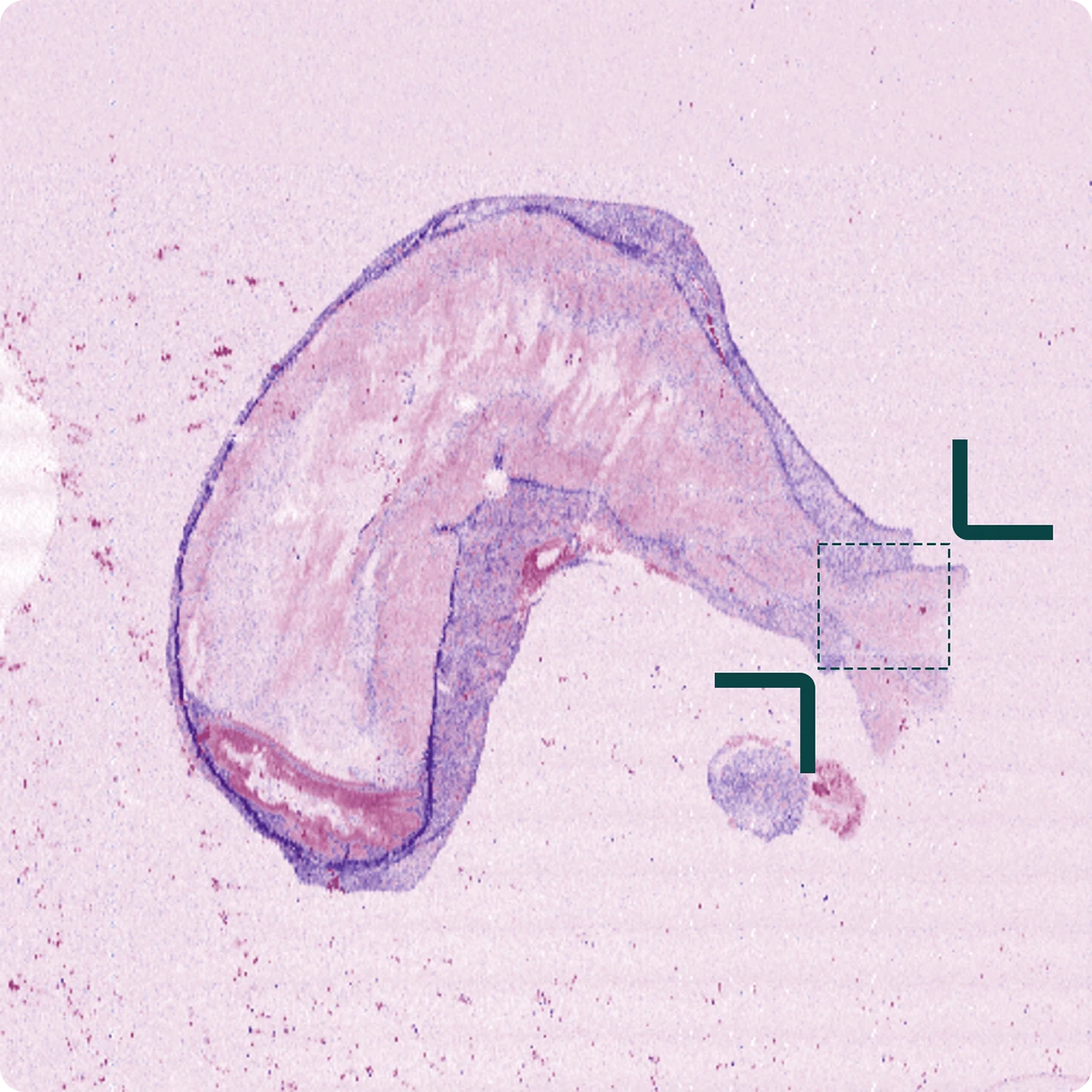
Macro scan or photo< 5 sec. |
Overview scan for orientation~min / cm2 |
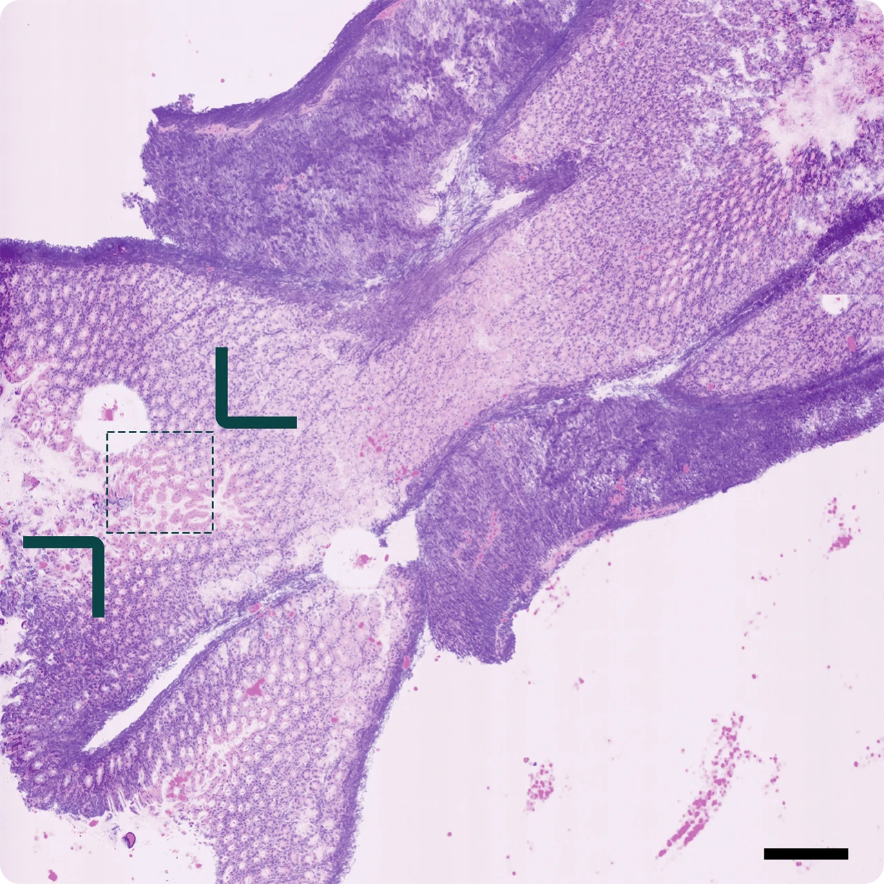
Detailed scan for diagnosis~mins / mm2 |
Intuitive sample navigation and multiscale scanning: Aquila combines a large imaging window with submicron spatial resolution, allowing users to navigate seamlessly from a macroscopic overview to cellular detail. Its core capability is the creation of virtual stains in minutes, transforming how tissue can be visualized and assessed directly from fresh, unstained samples. These digital images reproduce the familiar appearance of histological sections and can be reviewed immediately on site or remotely.
In addition, Aquila acquires multimodal optical signals based on Raman scattering, second harmonic generation, and two photon excited fluorescence, providing complementary molecular, structural, and functional information in the same dataset.
Layer insight
Three dimensional insight into tissue architecture: Aquila captures volume stacks through tens of micrometers of tissue, visualizing microstructures in context. The depth information clarifies layer organization and supports biopsy orientation and subsurface–surface correlation. [Sample courtesy of D. Kapsokalyvas (Maastricht University)]
How it works: Amplifying the molecular fingerprint with multimodal contrast
Every molecule carries a unique vibrational fingerprint that reveals its chemical identity. Aquila uses Raman-based imaging to capture this molecular information directly from fresh tissue, without stains or labels, and at imaging speeds suitable for clinical workflows. By combining molecular, structural, and functional contrast in a single scan, Aquila creates a rich, multimodal view of tissue architecture and composition.
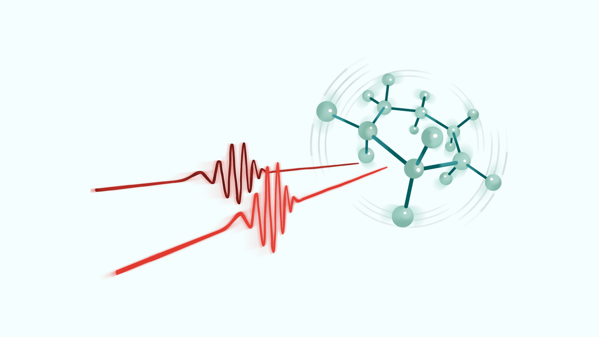
Molecular fingerprint: Mapping biochemical organization in its native context
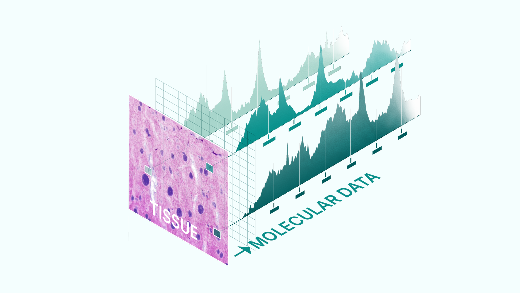
Aquila records Raman spectra per pixel to map proteins, lipids, nucleic acids, and carbohydrates label-free; these measurements expose morpho-molecular changes shaped by genetic and epigenetic factors, strengthening morphological assessment and informing intraoperative decisions and biomarker or AI-based analysis. This novel molecular data layer opens new opportunities for real-time insight into tumor biology and future biomarker discovery.
Digital integration: Integrated Aquila software suite
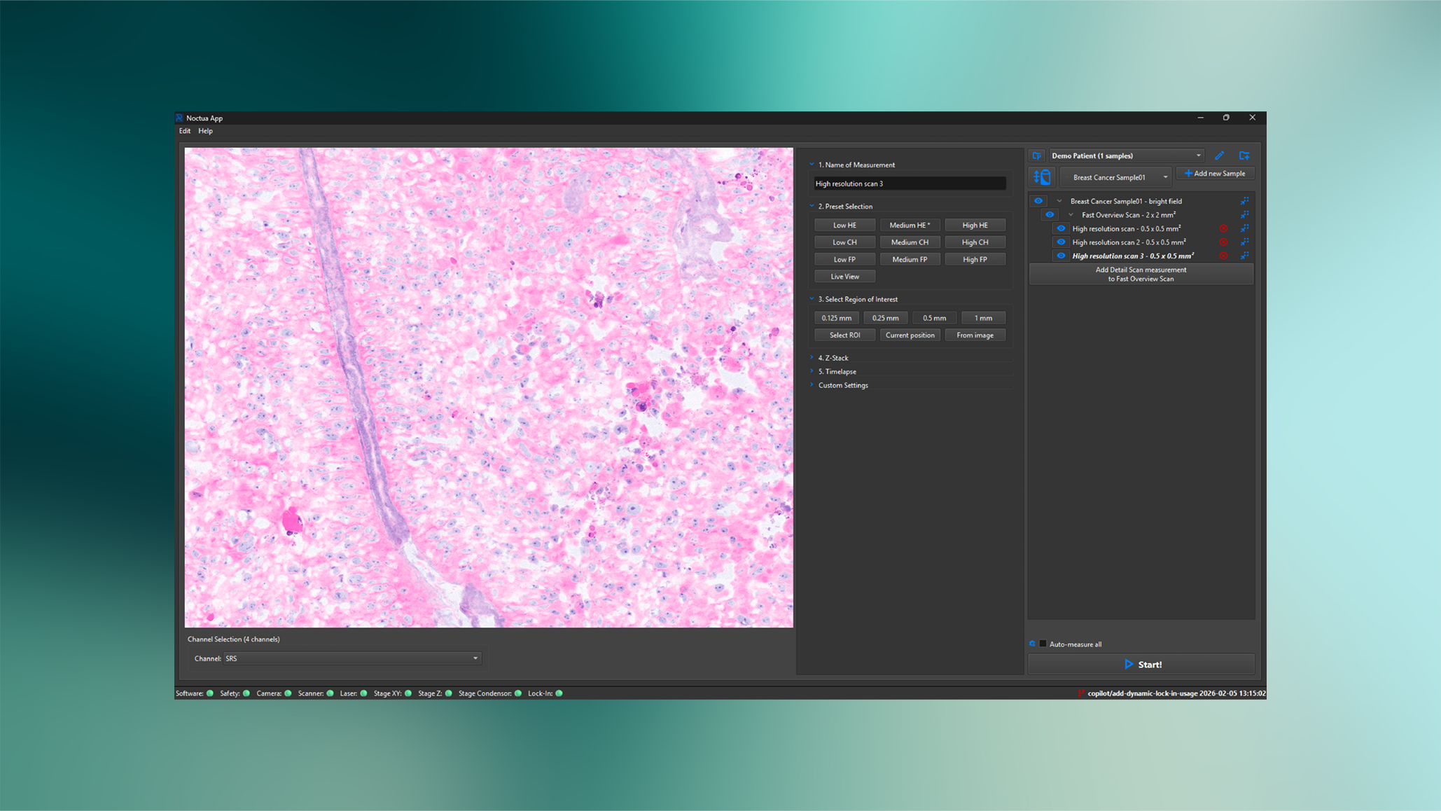
Aquila includes an intuitive software interface for image acquisition, visualization, and data management. Digital results can be reviewed immediately, stored securely, and exported in standard formats for further analysis or AI integration.
| File Name | Size | Link |
|---|---|---|
 Terms and Conditions V6 Terms and Conditions V6
|
207.42KiB | Download |